Diagnostic Hysteroscopy
Diagnostic hysteroscopy is a minimally invasive procedure that allows doctors to examine the inside of the uterus with precision. By providing a direct view of the uterine cavity, it helps identify conditions that may interfere with fertility, cause repeated pregnancy loss, or prevent successful embryo implantation. This technique has revolutionized fertility care, as it combines accuracy, safety, and sometimes immediate treatment in a single session.
Conditions Diagnosed
Diagnostic hysteroscopy is especially useful in identifying abnormalities that may remain hidden in other tests such as ultrasounds. It can detect:
- Uterine Polyps
- Small growths on the inner lining of the uterus that may interfere with embryo implantation or cause irregular bleeding.
- Fibroids Inside the Uterine Cavity (Submucosal Fibroids)
- Benign tumors that can distort the uterine cavity and reduce the chances of successful pregnancy.
- Early detection helps in planning removal before fertility treatments.
- Scar Tissue (Adhesions)
- Formed due to previous surgeries, infections, or miscarriages.
- Can prevent proper implantation or increase the risk of miscarriage if left untreated.
- Congenital Uterine Abnormalities
- Structural issues present from birth, such as a septum or bicornuate uterus, which may contribute to infertility or recurrent pregnancy loss.
By identifying these issues early, hysteroscopy allows doctors to intervene before they compromise fertility, giving patients the best chance of a healthy pregnancy.
Procedure Overview
Diagnostic hysteroscopy is generally quick, safe, and minimally uncomfortable:
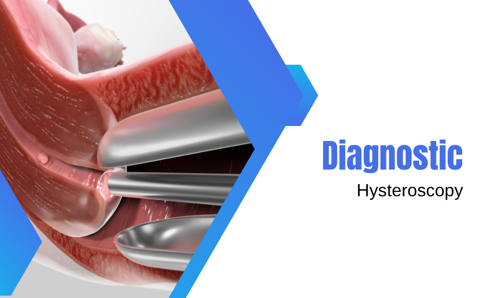
- A thin, lighted instrument called a hysteroscope is gently inserted through the cervix into the uterus.
- The hysteroscope is equipped with a camera, allowing real-time visualization of the uterine cavity on a monitor.
- In many cases, anesthesia is not required, though mild sedation or local anesthesia may be used for patient comfort.
- The procedure usually takes 10–20 minutes, after which patients can often resume normal activities the same day.
Some hysteroscopes also allow simultaneous treatment, such as removing polyps or small fibroids, making it a highly efficient diagnostic and therapeutic tool.
Benefits of Diagnostic Hysteroscopy
- Direct and Accurate Visualization
- Unlike imaging alone, hysteroscopy allows the doctor to see the uterine cavity directly, providing a definitive diagnosis.
- Detection of Hidden Abnormalities
- Small polyps, adhesions, or subtle congenital defects that may not appear on ultrasound can be easily identified.
- Combination of Diagnosis and Treatment
- Minor issues detected during the procedure can often be corrected immediately, avoiding additional procedures and reducing treatment time.
- Minimally Invasive and Safe
- No large incisions are needed, and the risk of complications is very low.
- Optimizes Fertility Outcomes
- Ensuring the uterine environment is healthy improves the chances of implantation and reduces the risk of miscarriage.
Role in Fertility Treatment
Dr. Abhilasha Mehta uses diagnostic hysteroscopy as a key step in preparing patients for fertility treatment, particularly in cases of:
- Recurrent pregnancy loss
- Unexplained infertility
- Abnormal uterine findings on imaging
By addressing hidden abnormalities and creating an optimal uterine environment, hysteroscopy significantly enhances the likelihood of a successful pregnancy. It is often combined with IVF cycles to maximize outcomes, ensuring that embryos are transferred into the healthiest possible environment.
In essence, diagnostic hysteroscopy provides clarity, precision, and actionable results, giving couples confidence that their fertility journey is based on accurate and thorough evaluation.
